RSNA 2013
Contents

| |||

|

|

| |
- The Slicer hands-on courses were sold out
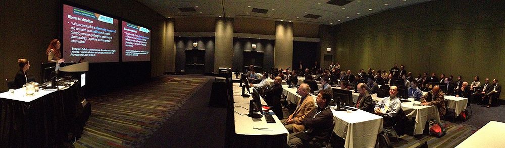
Quantitative Medical Imaging for Clinical Research and Practice
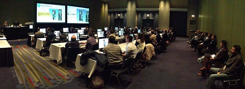
3D Interactive Visualization of DICOM Images for Radiology Applications


Agenda
| Sunday, December 1 | Monday, December 2 | Tuesday, December 3 | Wednesday, December 4 | Thursday, December 5 | Friday, December 6 |
|---|---|---|---|---|---|
| 8:00am-11:00am: 3D Slicer Exhibit: Quantitative Imaging Reading Room. Lakeside Learning Center Hall E, Exhibit LL-QRR3007. 11:00am-12:30pm: RSNA Refresher Course: "Quantitative Medical Imaging for Clinical Research and Practice: Hands-on Workshop." 12:30pm-1:30pm: Meet-The-Experts Session, 3D Slicer Exhibit: Quantitative Imaging Reading Room. 1:30pm-6:00pm: 3D Slicer Exhibit: Quantitative Imaging Reading Room. |
8:00am-11:00am: 3D Slicer Exhibit: Quantitative Imaging Reading Room. Lakeside Learning Center Hall E, Exhibit LL-QRR3007. 12:15pm-1:15pm: Meet-The-Experts Session, 3D Slicer Exhibit: Quantitative Imaging Reading Room. 1:15pm-6:00pm: 3D Slicer Exhibit: Quantitative Imaging Reading Room. |
8:00am-11:00am: 3D Slicer Exhibit: Quantitative Imaging Reading Room. Lakeside Learning Center Hall E, Exhibit LL-QRR3007. 12:30pm-2:00pm: RSNA Refresher Course: "3D Interactive Visualization of DICOM Images for Radiology Applications: Hands-on Workshop." 12:15pm-1:15pm: Meet-The-Experts Session, 3D Slicer Exhibit: Quantitative Imaging Reading Room. |
8:00am-12:15pm: 3D Slicer Exhibit: Quantitative Imaging Reading Room. Lakeside Learning Center Hall E, Exhibit LL-QRR3007. 12:15pm-1:15pm: Meet-The-Experts Session, 3D Slicer Exhibit: Quantitative Imaging Reading Room. 1:15pm-6:00pm: 3D Slicer Exhibit: Quantitative Imaging Reading Room. |
8:00am-12:15pm: 3D Slicer Exhibit: Quantitative Imaging Reading Room. Lakeside Learning Center Hall E, Exhibit LL-QRR3007. 12:15pm-1:15pm: 3D Slicer Exhibit: Quantitative Imaging Reading Room. 1:15pm-6:00pm: 3D Slicer Exhibit: Quantitative Imaging Reading Room. |
8:00am-12:45pm: 3D Slicer Exhibit: Quantitative Imaging Reading Room. Lakeside Learning Center Hall E, Exhibit LL-QRR3007. |
Introduction

3D Slicer will be featured at RSNA 2013 through a week-long series of events, which include two hands-on courses in RSNA classrooms and a 3D Slicer exhibit in the Quantitative Imaging Reading Room. The "Quantitative Medical Imaging for Clinical Research and Practice" course (Sonia Pujol, Katarzyna Macura and Ron Kikinis) has been selected by Radiology Today in the article "Challenges and Opportunities - A Sneak Peek at RSNA 2013 With Views From President Sarah S. Donaldson, MD" Radiology Today Vol. 14 No. 10 P. 24" in two categories 'Finance and Support Research in Radiologic Sciences' and 'Promote and Advance Precision Radiology Without Increasing Costs'.
Quantitative Medical Imaging for Clinical Research and Practice

Technological breakthroughs in medical imaging hardware and the emergence of increasingly sophisticated image processing software tools permit the visualization and display of complex anatomical structures with increasing sensitivity and specificity. This course begins with an introductory presentation of state-of-the-art, clinical examples of quantitative imaging biomarkers for diagnosis and clinical trial outcome measures. Cases from multiple imaging modalities and from multiple organ systems will be highlighted to illustrate the depth and breath of this field. Participants will then be led through a series of tutorials on the basics of viewing and processing DICOM volumes in 3D using 3D SLICER (www.slicer.org). The version of the software that will be used during this course is Slicer4.3.1-1 and it can be downloaded here. A hands-on Quantitative Imaging tutorial ppt with pre-computed datasets will focus on basic use of 3D Slicer software, quantitative measurements from PET/CT studies, and volumetric analysis of meningioma. The course is offered in collaboration with the Johns Hopkins Institute for Clinical and Translational Research. For additional training materials on the software, please visit the 3D Slicer Compendium.
- CME Credit Categories: Biomarkers/Quantitative Imaging; Informatics
- AMA PRA Category 1 Credits™: 1.5
- ARRT Category A+ Credit: 1.5
Teaching Faculty
- Sonia Pujol, Ph.D., Surgical Planning Laboratory, Harvard Medical School, Department of Radiology, Brigham and Women’s Hospital, Boston MA.
- Katarzyna J. Macura MD, PhD., Department of Radiology and Radiological Sciences, The Johns Hopkins Medical Institutions, Baltimore, MD.
- Ron Kikinis, M.D., Surgical Planning Laboratory, Harvard Medical School, Department of Radiology, Brigham and Women’s Hospital, Boston MA.
Logistics
- Date: Sunday December 1st; 11:00 am-12:30 pm
- Location: Room TBA , McCormick Conference Center, Chicago, IL.
3D Interactive Visualization of DICOM images for Radiology Applications

This course will be offered by the National Alliance for Medical Image Computing (NA-MIC) in conjunction with the Neuroimage Analysis Center (NAC), at the 99th Scientific Assembly and Annual Meeting of the Radiological Society of North America (RSNA 2013). As part of the outreach missions of these NIH funded National Centers, we have developed an offering of freely available, multi-platforms open source software to enable medical image analysis research. The 3D Visualization course ppt along with the 3D Visualization-part1 and 3D Visualization-part2 datasets aim to introduce translational clinical scientists to the basics of viewing and interacting in 3D with DICOM volumes and anatomical models using the 3DSlicer software. The version of the software that will be used during this course is Slicer4.3.1-1 and it can be downloaded here. For additional training materials on the software, please visit the 3D Slicer Compendium.
- CME Content Code: Informatics
- CME Credits Categories:
- AMA PRA Category 1 Credits™: 1.5
- ARRT Category A+ Credit: 1.5
Teaching Faculty
- Sonia Pujol, Ph.D., Surgical Planning Laboratory, Harvard Medical School, Department of Radiology, Brigham and Women’s Hospital, Boston MA.
- Ron Kikinis, M.D., Surgical Planning Laboratory, Harvard Medical School, Department of Radiology, Brigham and Women’s Hospital, Boston MA.
- Kitt Shaffer, M.D. Ph.D., Boston University School of Medicine, Department of Radiology, Boston Medical Center, Boston, MA.
Logistics
- Date: Tuesday December 3; 12:30-2:00pm
- Location: Room TBA McCormick Conference Center, Chicago, IL.
Quantitative Imaging Reading Room

3DSlicer: An open-source platform for segmentation, registration, quantitative imaging and 3D visualization of multi-modal image data. The 3DSlicer exhibit will consist in a series of thematic demonstrations using multi-modal image datasets, which include MRI, CT, PET and DCE. A team of 3DSlicer experts will be running the demonstrations with sample datasets or, where appropriate, data provided by attendees, and will deliver a series of daily 60-minute Meet-the-Experts sessions. The hands-on sessions will demonstrate 3D visualization functionalities (e.g. volume rendered head, thoracic and abdominal CT scans, surface rendered atlases of brain, knee, and abdomen), segmentation tools (e.g. semi-automated segmentation of a glioma case, MRI-based automated parcellation of the human brain), quantitative imaging features (e.g. longitudinal analysis of meningioma growth, PET/CT quantitative assessment of tumor response, traumatic brain injury cases follow-up) as well as image-guided therapy applications (e.g. exploration of peritumoral white matter for neurosurgical planning, registration of prostate imaging data). In addition, the exhibit will present new workflows for full brain tractography and multi-volume exploration of prostate DCE MRI data.
Teaching Faculty
- Sonia Pujol, Ph.D., Surgical Planning Laboratory, Harvard Medical School, Department of Radiology, Brigham and Women’s Hospital, Boston MA.
- Ron Kikinis, M.D., Surgical Planning Laboratory, Harvard Medical School, Department of Radiology, Brigham and Women’s Hospital, Boston MA.
- Steve Pieper, Ph.D., Isomics Inc.
- Jayender Jagadeesan, Ph.D., Surgical Planning Laboratory, Harvard Medical School, Department of Radiology, Brigham and Women’s Hospital, Boston MA.
- Alireza Mehrtash, M.Sc., Surgical Planning Laboratory, Harvard Medical School, Department of Radiology, Brigham and Women’s Hospital, Boston MA.
Logistics
- Date: Sunday December 1st -Friday December 6; 8:00am-5:00pm
- Location: Exhibit LL-QRR3007, Lakeside Learning Center, McCormick Conference Center, Chicago, IL.
Meet the Experts Sessions
Daily one-hour Meet-The-Experts sessions will be held from Sunday to Thursday as part of the Quantitative Imaging Room exhibit. The sessions will give participants an opportunity to interact with the on-site faculty through software demonstrations and general question and answer sessions, which will include a series of hands-on demonstrations of the 3DSlicer modules for quantitative imaging.
- Faculty:
- Ron Kikinis, MD
- Sonia Pujol, PhD
- Steve Pieper, PhD
- Jayender Jagadeesan, Ph.D.
- Alireza Mehrtash, M.Sc.
- Schedule:
- Sunday, December 1: 12:30 p.m. – 1:30 p.m.
- Monday, December 2: 12:15 p.m. – 1:15 p.m.
- Wednesday, December 4: 12:15 p.m. – 1:15 p.m.
- Thursday, December 5: 12:15 p.m. – 1:15 p.m.